
What you need to know
Hackensack University Medical Center (HUMC) is the first center in New Jersey and the second in the world to use an imaging technology before surgery to assess living donor kidneys more precisely and comprehensively than conventional imaging techniques, as well as during surgery to help guide the removal of donor kidneys.
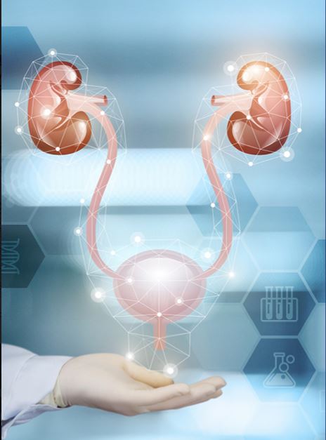
HUMC’s living donor kidney surgeons are using the imaging software in conjunction with CT scanning to create virtual 3D models of the kidney that assist with preoperative planning as well as intraoperative navigation during robotic-assisted donor nephrectomy.
The software, called IRIS, enables HUMC surgeons to use kidneys that might have been deemed unfit for transplant based solely on the findings of standard CT scanning, potentially expanding the pool of donor kidneys available to patients.
The virtual anatomical 3D digital models generated by IRIS — which can be downloaded to any iOS device, such as an iPhone or iPad — can be rotated and manipulated by the surgeon to see any part of the kidney in detail, including the exact location of arteries, veins, and other structures. Using IRIS can make the removal of a donor kidney faster, more efficient, and safer because the surgeon has more information before and during the surgical procedure.
Since June 2020, HUMC living donor kidney surgeons have used IRIS technology in more than 50 patients, and this has now become a routine part of their preoperative assessment.
In the foreseeable future, HUMC's living donor kidney team have a goal of utilizing IRIS assistance to help remove donor kidneys using single-port robotic surgery, removing the kidney robotically through a single small incision.